At Pomona Valley Hospital Medical Center, we design our cardiac diagnostic
services to provide accurate assessments and personalized care for patients
with cardiovascular conditions. Our knowledgeable cardiologists and imaging
specialists use advanced technologies to give our patients thorough evaluations
and the right treatment plan.
Diagnostic Procedures:
Coronary CT (Computed Tomography)
Cardiovascular Imaging
CT or CAT scan
Coronary CT (Computed Tomography)
Coronary CT, also known as computed tomography angiography (CTA), is a
noninvasive imaging technique that uses advanced CT technology to provide
detailed pictures of the coronary arteries and heart structures. This
procedure allows our cardiologists to assess the presence of coronary
artery disease, detect blockages and evaluate the general health of the
heart and blood vessels. Coronary CT helps identify calcifications, plaque
buildup, and narrowing of the arteries, providing vital information for
treatment decisions and risk assessment.
Cardiovascular Imaging
X-rays are a form of radiation, like light or radio waves, that is focused
into a beam of light. However, unlike a beam of light, X-rays can pass
through most objects, including the human body.
When X-rays strike a photographic film, they can produce a picture. Dense
tissues in the body, such as bones, block (absorb) many X-rays and appear
white on an X-ray picture. Less dense tissues, such as muscles and organs,
block fewer X-rays (more X-rays pass through) and appear in shades of
gray. X-rays that pass only through air appear black on an X-ray picture.
CT or CAT scan
A computed tomography (CT) scan uses X-rays to make detailed pictures of
structures inside the body.
During the test, you will lie on a table hooked to the CT scanner, a large
doughnut-shaped machine. The CT scanner sends X-ray pulses through the
body. Each pulse lasts less than a second and takes a picture of a thin
slice of the organ or area. One part of the scanning machine can tilt
to take photos from different positions and save them on a computer.
A CT scan can study any body organ, such as the liver, pancreas, intestines,
kidneys, adrenal glands, lungs, and heart. It also can study blood vessels,
bones, and the spinal cord.
An iodine dye (contrast material) is used to make structures and organs
easier to see on CT pictures. The dye may be used to check blood flow,
find tumors, and look for other problems. It can be put in a vein (IV)
in your arm, or you may drink it for some tests. CT pictures may be taken
before and after the dye is used.
At Pomona Valley Hospital Medical Center, precision and accuracy in cardiac
diagnostic tests and procedures. We leverage advanced imaging technologies
and expert interpretation by our cardiologists and imaging specialists.
Our commitment to excellence ensures that patients receive thorough evaluations,
timely diagnoses and optimal care plans tailored to their cardiovascular needs.

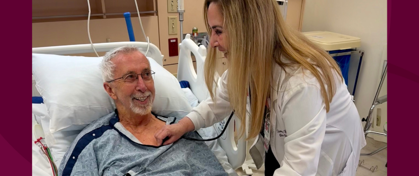
 July 23 Life After Stroke, New Beginnings

[1].png)
.png)


.png)
.png)