At Pomona Valley Hospital Medical Center, we strongly emphasize accuracy
and precision in vascular diagnostics by utilizing forefront imaging technology
and providing knowledgeable interpretation from our team of qualified
imaging specialists and vascular specialists. Our dedication to quality
guarantees that patients receive in-depth assessments, prompt diagnosis,
and ideal treatment regimens. Our committed team offers thorough evaluations
and individualized care to support your vascular well-being, whether you're
worried about varicose veins, deep vein thrombosis, or other vascular
conditions.
Vein Mapping for an Arterial-Venous Fistula for Dialysis (AVP)
Pneumogram
Venous Duplex
The imaging method known as venous duplex ultrasound uses high-frequency
sound waves, or ultrasound, to produce images of the veins that carry
blood back to the heart. Leg and ankle veins are inspected during a lower
extremity venous duplex scan. The two components of a venous duplex ultrasound
exam are the Doppler and the two-dimensional echo, which are performed
simultaneously. Images of the veins under examination are produced by
the 2-D echo component. The Doppler element generates audible noises that
aid a medical professional in determining the direction and speed of blood
flow through veins.
Pulmonary Angiogram
An angiogram of the lung is an X-ray test that uses fluoroscopy to take
pictures of the blood flow within the lung's blood vessels. A thin, flexible
tube called a catheter is usually placed into a blood vessel in the groin
or just above the elbow and guided through the heart to the lungs. Then,
a dye (contrast material) containing iodine is injected into the vessel
being studied to make it more visible in the X-ray pictures.
A lung (pulmonary) angiogram evaluates the arteries leading to the lungs
(pulmonary arteries) and the blood vessels within the lungs. It can also
detect narrowing or a blockage in a blood vessel that slows or prevents
blood flow. Angiogram pictures can be produced on regular X-ray films
or stored as digital images in a computer.

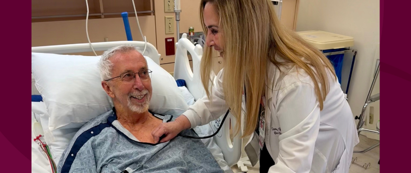
 July 23 Life After Stroke, New Beginnings

[1].png)
.png)


.png)
.png)